|
Listen to this article
|

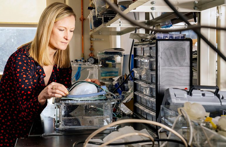
Ellen Roche with the soft, implantable ventilator designed by her and her team. | Source: MIT, M. Scott Brauer
Researchers at MIT have designed a soft, robotic implantable ventilator that can augment the diaphragm’s natural contractions.
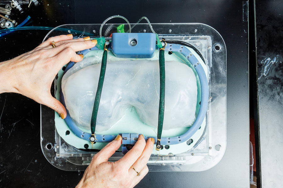
The implantable ventilator is made from two soft, balloon-like tubes that would be implanted to lie over the diaphragm. When inflated with an external pump, the tubes act as artificial muscles that push down the diaphragm and help the lungs expand. The tubes can be inflated to match the diaphragm’s natural rhythm.
The diaphragm lies just below the ribcage. It pushes down to create a vacuum for the lungs to expand into so they can draw air in, and then relaxes to let air out.
The tubes in the ventilator are similar to McKibben actuators, a kind of pneumatic device. The team attached the tubes to the ribcage at either side of the diaphragm, so that the device was laying across the muscle from front to back. Using a thin external airline, the team connected the tubes to a small pump and control system.
This soft ventilator was designed by Ellen Roche, an associate professor of mechanical engineering and member of the Institute for Medical Engineering and Science at MIT and her colleagues. The research team created a proof-of-concept design for the ventilator.
“This is a proof of concept of a new way to ventilate,” Roche told MIT News. “The biomechanics of this design are closer to normal breathing, versus ventilators that push air into the lungs, where you have a mask or tracheostomy. There’s a long road before this will be implanted in a human. But it’s exciting that we could show we could augment ventilation with something implantable.”
According to Roche, the key to maximizing the amount of work the implantable pump does is by giving the diaphragm an extra push downwards when it naturally contracts. This means the team didn’t have to try to mimic exactly how the diaphragm moves, just create a device that is capable of giving that push.

The implantable ventilator is made from two tubes that lay across the diaphragm. | Source: MIT
Roche and her team tested the system on anesthetized pigs. After implanting the device, they monitored the pigs’ oxygen levels and used ultrasound imaging to observe diaphragm function. Generally, the team found that the ventilator increased the amount of air that the pigs’ lungs could draw in with each breath. The device worked best when the contractions of the diaphragm and the artificial muscles were working in sync, allowing the pigs’ lungs to bring in three times the amount of air they could without assistance.
The team hopes that its device could help people struggling with chronic diaphragm dysfunctions, which can be caused by ALS, muscular dystrophy and other neuromuscular diseases, paralysis and damage to the phrenic nerve.
The research team included Roche, a former graduate student at MIT Lucy Hu, Manisha Singh, Diego Quevedo Moreno, Jean Bonnemain of Lausanne University Hospital in Switzerland and Mossab Saeed and Nikolay Vasilyev of Boston Children’s Hospital.


Tell Us What You Think!